Introduction
Since the first case was reported in December 2019, COVID-19 has rapidly evolved into a global crisis, resulting in more than 1 million deaths worldwide and becoming the deadliest flu pandemic since the Spanish flu in 1918. Governments and healthcare systems in almost every country are taking extreme measures to contain the infection, including strict curfews and travel restrictions. These public health measures have severely impacted the global economy, pushing the United Kingdom into the worst recession on record in the first two quarters of 2020 and driving almost 200,000 people into redundancies.
The acute symptoms of COVID-19, or severe acute respiratory syndrome coronavirus 2 (SARS-CoV-2), that have been characterized to date are generally consistent with other upper respiratory infections and include fever, cough, shortness of breath, and chest pain. However, as the global scientific community begins to understand the syndromic complexity of the disease, there is increasing evidence to suggest that COVID-19 is more than a respiratory syndrome, with lasting effects on other tissues, such as the kidneys,[1] liver,[2] heart,[3] and nervous system.
Mild neurological symptoms and long COVID
Various neurological abnormalities have been described in patients with COVID-19, ranging from mild to fatal and involving both the central and peripheral nervous systems. The exact prevalence of patients experiencing neurological problems following a COVID-19 infection varies, but one report states that up to 80% of patients admitted to hospital with COVID-19 suffer from at least one neurological symptom.[12] In patients with mild COVID‑19, the neurological effects are mostly limited to non-specific symptoms, such as crippling fatigue, dizziness, muscle, and joint pain, headaches, and loss of smell and taste, which are not dissimilar to those routinely observed in other respiratory virus infections like the flu. However, unlike the flu, these mild neurological symptoms become chronic in about half of these patients, lasting for weeks or even months after apparent recovery from the respiratory syndrome.[4] A new term has been coined by both experts and the public alike to describe this cohort—the “long COVID” sufferers.
Medical anomalies and brainstem damage
While the chronic neurological symptoms of long COVID can be life-changing, they are often not fatal, unlike symptoms in patients suffering from severe COVID-19. A mystery currently baffling doctors and scientists is a phenomenon known as “happy hypoxia” or, perhaps more accurately, “silent hypoxia.” In healthy individuals, blood oxygen saturation levels should be above 95%. Saturation levels below 80% may result in impairment of mental function, and levels below 75% typically cause a loss of consciousness. However, there are now increasing reports of patients being conscious and comfortable, with no breathing difficulties, while having saturation levels of less than 75%. There is even a report of a patient with a saturation level of 60% talking on a mobile phone with no apparent discomfort.[5] While the exact mechanism behind this phenomenon is unknown, there has been speculation that it is a result of the virus attacking the brainstem.[6] The brainstem is a small structure at the base of the brain responsible for regulating cardiac and respiratory functions including heart rate and breathing rate. Thus, damage to the brainstem can seriously affect one’s ability to regulate breathing. Scientists also believe that brainstem damage could be the cause of severe respiratory failure in some COVID-19 patients.[7]
Ischemic stroke
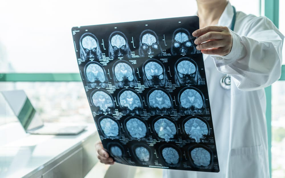
Beyond the brainstem, there is mounting evidence that the neurological effects of COVID-19 may be more widespread. A magnetic resonance imaging study conducted in 126 patients showed that only 15% of patients did not have any abnormalities in their brain images, and up to 25% exhibited blocked or narrowed blood vessels in the brain.[8] In addition, a multicenter study reported that up to 88% of patients exhibited abnormally increased tendencies toward blood clotting.[21] In line with these findings, a paper recently reported that the SARS-CoV-2 virus is seven times more likely to result in ischemic strokes, caused by blood clots in the brain, compared to normal flu.[9] There are also increasing numbers of case studies that demonstrate the ability of the virus to invade the brain directly, causing encephalitis (inflammation of the brain) or meningitis (inflammation of the membranes that surround the brain and spinal cord).[10][11]
How does COVID reach the brain?
While scientists do not yet have sufficient evidence to conclusively determine the entry mechanism of the virus to the brain, there are some working hypotheses based on studies relating to the discovery of viral particles in the brain and/or spinal fluid of COVID-19 patients.
Loss of smell is one of the most frequent neurological manifestations of COVID-19, and brain imaging studies have provided evidence that the olfactory cortex in the brain can be infected by the virus.[15] Once in the olfactory cortex, the virus can, in theory, spread easily to other regions of the brain.
Another common entry route is via the blood-brain barrier (BBB). The barrier is made up of a tightly packed layer of cells that line the blood vessels in the brain and the spinal cord. It can prevent large molecules and disease-causing organisms such as bacteria and viruses from entering the central nervous system through the bloodstream. Factors like age and pre-existing cardiovascular and/or neurological disease can damage this barrier, increasing the likelihood of the virus entering the brain. It has also been discovered that a number of SARS-CoV-2-associated small proteins, such as interleukin (IL)-6, IL-1β, tumor necrosis factor (TNF), and IL-17, have the ability to disrupt the BBB, which can also facilitate the entry of the virus.[16]
Indirect mechanisms of neurological damage
The indirect effects of COVID-19 on the brain are closer to those of other viral infections such as influenza and glandular fever. As the virus targets the respiratory system, other organs are often caught in the “crossfire.” Lung damage and respiratory failure in severely ill patients can seriously decrease the amount of oxygen available to the brain, resulting in brain damage. This is supported by autopsy studies in COVID-19 patients; damage is reported in brain regions most susceptible to oxygen deprivation, showing a definite link between the brain and the lungs.[17–19]
The brain can also be damaged as a result of a “cytokine storm,” which is the uncontrolled and excessive release of cytokines. The precise biological mechanism for this is not entirely understood, but it may be caused by an exaggerated response when the immune system encounters a new and highly pathogenic invader. Some causative agents include viral respiratory infections, Epstein–Barr virus, cytomegalovirus, group A streptococcus, and non-infectious conditions such as graft-versus-host disease. This out-of-proportion and out-of-place release of cytokines are usually destructive for all vital organs. For the brain, this becomes extremely devastating and further paves the way for a variety of inflammatory diseases like meningitis, encephalitis, and meningoencephalitis.[20]
Challenges in tracking neurological symptoms
It has been difficult to determine the extent and severity of these neurological issues for two main reasons. First, many of the mild neurological symptoms can be vague and difficult to recognize. For lots of patients, symptoms like fatigue, dizziness, and confusion are just part of “getting ill.” Additionally, more severe symptoms can be missed by physicians if they do not know what to look out for. For example, someone experiencing a seizure may just appear dazed, and sedation in an ICU environment may mask neurological symptoms in the most severely ill cohort.
Second, many people suffering from COVID-19 are never actually tested for the virus, especially if they do not have any respiratory symptoms or a fever. A meta-analysis conducted over 41 studies with more than 50,000 patients showed that up to 16% of people who test positive for COVID-19 do not display any symptoms,[13] while a release by the WHO suggests that up to 80% of infections are mild or asymptomatic.[14] However, in some countries, due to limited resources, mass testing of the population is not possible. For example, the United Kingdom’s National Health Service only provides free COVID-19 tests if at least one of the following symptoms are present: high temperature, continuous cough, or loss/change in sense of smell or taste. As such, it is not possible to accurately account for COVID-19 infection rates, further challenging the tracking of COVID-19-associated neurological symptoms.
Moving forward
COVID-19 remains a new disease to all of us, and despite unprecedented international efforts to characterize it, there is still a long list of unanswered questions about the neurological implications and a much shorter list of solutions.
For patients suffering from long COVID, there has been increased recognition and awareness from the medical community. The British Medical Journal recently published guidelines about delivering primary care to these patients[22]; measures proposed include providing resources on fatigue management and pulse oximeters that monitor potential silent hypoxia episodes. There are also various support groups set up by national and local governments to help patients cope with the neurological and psychological symptoms of long COVID.
For the more severe neurological symptoms of COVID-19, a set of treatment guidelines has yet to be published. There are simply too many unknowns about the pathophysiology of this disease to say for certain what might be the best way to prevent and treat the neurological symptoms. As increasing numbers of studies involving brain imaging and cognitive testing are conducted on COVID-19 patients, and with a clearer understanding of the neurological effects, it will be feasible to develop comprehensive guidance for doctors and patients and identify therapeutics that could prevent neurological damage caused by COVID-19.
References
[1] Hirsch JS, Ng JH, Ross DW, et al. Acute kidney injury in patients hospitalized with COVID-19. Kidney Int. 2020;98(1):209-218. doi:10.1016/j.kint.2020.05.006
[2] Alqahtani SA, Schattenberg JM. Liver injury in COVID-19: The current evidence. United Eur Gastroenterol J. 2020;8(5):509-519. doi:10.1177/2050640620924157
[3] Tomasoni D, Italia L, Adamo M, et al. COVID-19 and heart failure: from infection to inflammation and angiotensin II stimulation. Searching for evidence from a new disease. Eur J Heart Fail. 2020;22(6):957-966. doi:10.1002/ejhf.1871
[4] Townsend L, Dyer AH, Jones K, et al. Full Title: Persistent fatigue following SARS-CoV-2 infection is common and independent of severity of initial infection Short title: Fatigue following SARS-CoV-2 infection. medRxiv. July 2020:2020.07.29.20164293. doi:10.1101/2020.07.29.20164293
[5] Tobin MJ, Laghi F, Jubran A. Why COVID-19 silent hypoxemia is baffling to physicians. Am J Respir Crit Care Med. 2020;202(3):356-360. doi:10.1164/rccm.202006-2157CP
[6] Soliz J, Schneider-Gasser EM, Arias-Reyes C, et al. Coping with hypoxemia: Could erythropoietin (EPO) be an adjuvant treatment of COVID-19? Respir Physiol Neurobiol. 2020;279:103476. doi:10.1016/j.resp.2020.103476
[7] Manganelli F, Vargas M, Iovino A, Iacovazzo C, Santoro L, Servillo G. Brainstem involvement and respiratory failure in COVID-19. Neurol Sci. 2020;41(7):1663-1665. doi:10.1007/s10072-020-04487-2
[8] Gulko E, Oleksk ML, Gomes W, et al. MRI Brain Findings in 126 Patients with COVID-19: Initial Observations from a Descriptive Literature Review. Am J Neuroradiol. September 2020. doi:10.3174/ajnr.a6805
[9] Merkler AE, Parikh NS, Mir S, et al. Risk of Ischemic Stroke in Patients with Coronavirus Disease 2019 (COVID-19) vs Patients with Influenza. JAMA Neurol. 2020. doi:10.1001/jamaneurol.2020.2730
[10] Ye M, Ren Y, Lv T. Encephalitis as a clinical manifestation of COVID-19. Brain Behav Immun. 2020;88:945-946. doi:10.1016/j.bbi.2020.04.017
[11] Moriguchi T, Harii N, Goto J, et al. A first case of meningitis/encephalitis associated with SARS-Coronavirus-2. Int J Infect Dis. 2020;94:55-58. doi:10.1016/j.ijid.2020.03.062
[12] Liotta EM, Batra A, Clark JR, et al. Frequent neurologic manifestations and encephalopathy‐associated morbidity in Covid‐19 patients. Ann Clin Transl Neurol. October 2020:acn3.51210. doi:10.1002/acn3.51210
[13] He J, Guo Y, Mao R, Zhang J. Proportion of asymptomatic coronavirus disease 2019: A systematic review and meta-analysis. J Med Virol. August 2020:jmv.26326. doi:10.1002/jmv.26326
[14] Coronavirus disease (COVID-19): Similarities and differences with influenza. https://www.who.int/emergencies/diseases/novel-coronavirus-2019/question-and-answers-hub/q-a-detail/q-a-similarities-and-differences-covid-19-and-influenza. Accessed October 25, 2020.
[15] Lu Y, Li X, Geng D, et al. Cerebral Micro-Structural Changes in COVID-19 Patients – An MRI-based 3-month Follow-up Study: A brief title: Cerebral Changes in COVID-19. EClinicalMedicine. 2020;25. doi:10.1016/j.eclinm.2020.100484
[16] Iadecola C, Anrather J, Kamel H. Effects of COVID-19 on the Nervous System. Cell. 2020;183(1):16-27.e1. doi:10.1016/j.cell.2020.08.028
[17] Kantonen J, Mahzabin S, Mäyränpää MI, et al. Neuropathologic features of four autopsied COVID-19 patients. Brain Pathol. August 2020:bpa.12889. doi:10.1111/bpa.12889
[18] Reichard RR, Kashani KB, Boire NA, Constantopoulos E, Guo Y, Lucchinetti CF. Neuropathology of COVID-19: a spectrum of vascular and acute disseminated encephalomyelitis (ADEM)-like pathology. Acta Neuropathol. 2020;140(1):1-6. doi:10.1007/s00401-020-02166-2
[19] Solomon IH, Normandin E, Bhattacharyya S, et al. Neuropathological Features of Covid-19. N Engl J Med. 2020;383(10):989-992. doi:10.1056/NEJMc2019373
[20] Mishra R, Banerjea AC. Neurological Damage by Coronaviruses: A Catastrophe in the Queue! Front Immunol. 2020;11:2204. doi:10.3389/fimmu.2020.565521
[21] Helms J, Tacquard C, Severac F, et al. High risk of thrombosis in patients with severe SARS-CoV-2 infection: a multicenter prospective cohort study. Intensive Care Med. 2020;46(6):1089-1098. doi:10.1007/s00134-020-06062-x
[22] Greenhalgh T, Knight M, A’Court C, Buxton M, Husain L. Management of post-acute covid-19 in primary care. BMJ. 2020;370. doi:10.1136/bmj.m3026

Belinda Ding
Scientific Consultant
Belinda Ding is a scientific consultant at Biochromex. She is a PhD-level Clinical Neuroscientist at the University of Cambridge. Her research focuses on developing cutting-edge MRI techniques for ultra-high field imaging.
